Cutting-Edge Instrumentation
Cutting-Edge Instrumentation
Precision tools powering advanced research, innovation, and structural characterization.
At the heart of the Hephaestus Laboratory lies an unparalleled infrastructure. Representing a strategic investment in scientific excellence, our facility houses an impressive array of specialized equipment designed for rigorous physical, chemical, and structural analysis. Explore our state-of-the-art instruments, meticulously maintained to bridge the gap between theoretical research and real-world industrial applications.
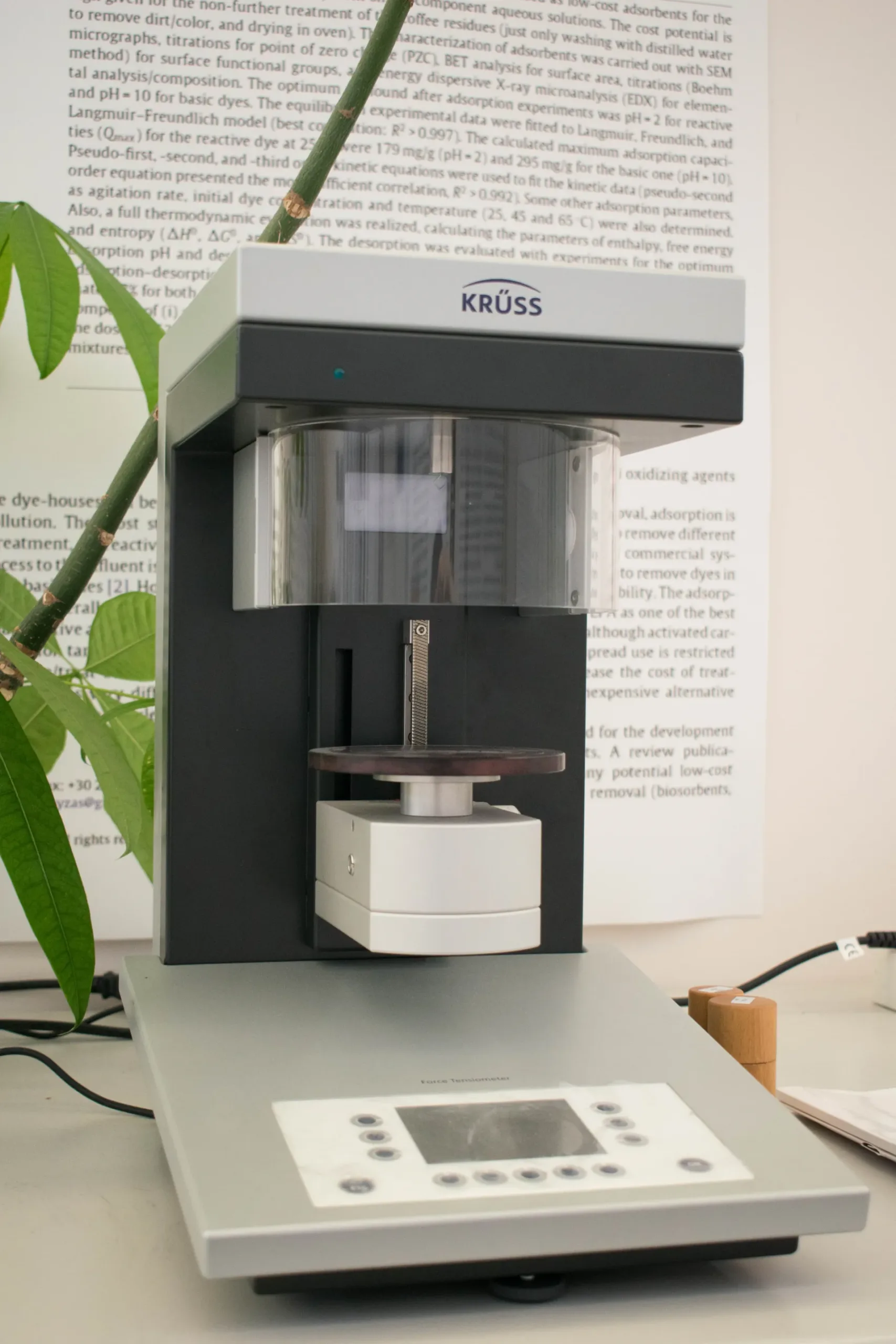
Optical tensiometer / contact angle meter — Theta Lite, Biolin Scientific
Instrument for measuring the wettability and surface energy of solid materials through static and dynamic contact angle analysis. A controlled liquid droplet is deposited on a sample surface, and high-resolution imaging software calculates the angle formed at the solid–liquid–air interface. The system is useful for evaluating surface treatments, coatings, adhesion behavior, hydrophobicity/hydrophilicity, pharmaceutical coatings, cosmetic formulations, and material–liquid interactions. Automated dosing and image analysis improve reproducibility and allow rapid comparison of different surfaces.
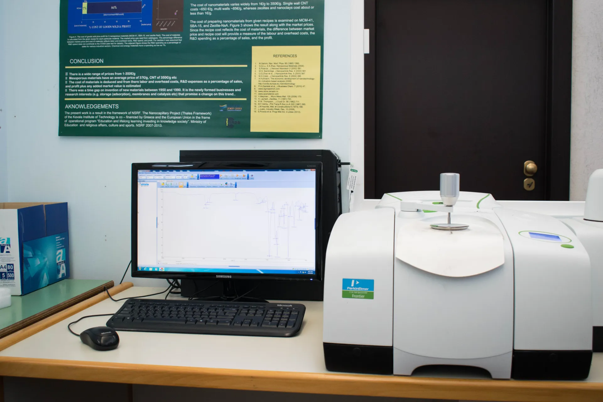
ATR-FTIR spectrometer — Frontier ATR-FTIR, PerkinElmer
Fourier-transform infrared spectroscopy system equipped with attenuated total reflectance capability for rapid chemical identification of solids, liquids, polymers, organic compounds, and some inorganic materials. The ATR accessory allows direct analysis with minimal sample preparation, avoiding pellet preparation or complex pretreatment. The instrument records infrared absorption spectra that reveal characteristic molecular vibrations and functional groups. It is used for material identification, polymer characterization, pharmaceutical analysis, quality control, forensic analysis, and monitoring chemical changes in research samples.
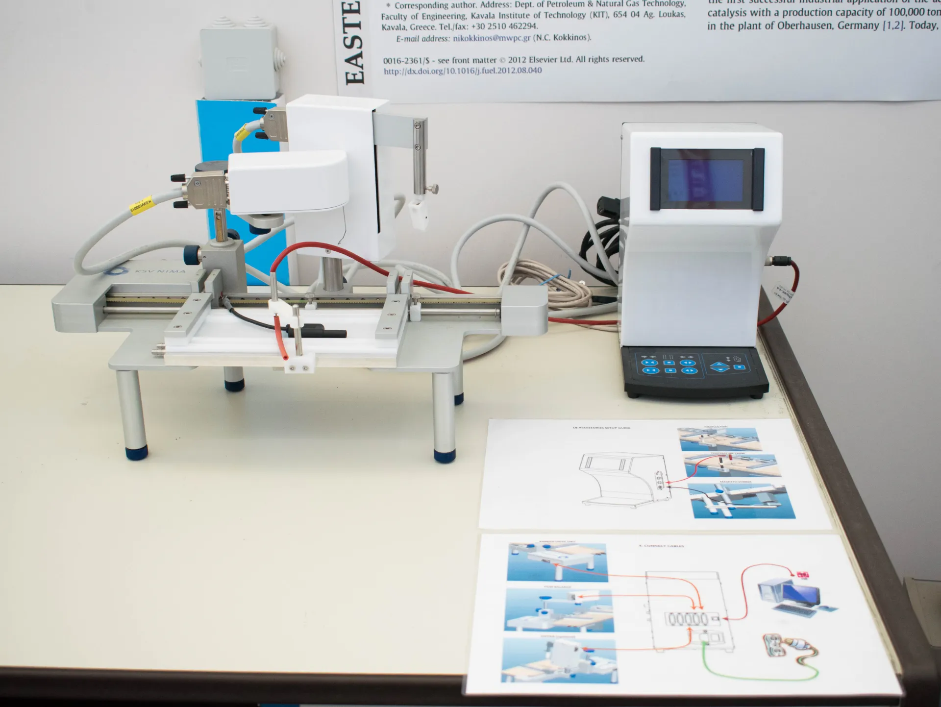
Langmuir–Blodgett trough — KSV NIMA Langmuir-Blodgett system
Instrument for preparing controlled monolayers and multilayer ultrathin films at the air–water interface and transferring them onto solid substrates. Molecules, polymers, lipids, or nanoparticles are spread on a liquid subphase and compressed using movable barriers while surface pressure is monitored. The system enables precise control of molecular packing, surface pressure, deposition speed, and film transfer. It is used for fabricating organized thin films for nanotechnology, sensors, biomimetic coatings, organic electronics, photonics, and surface-functionalized materials.
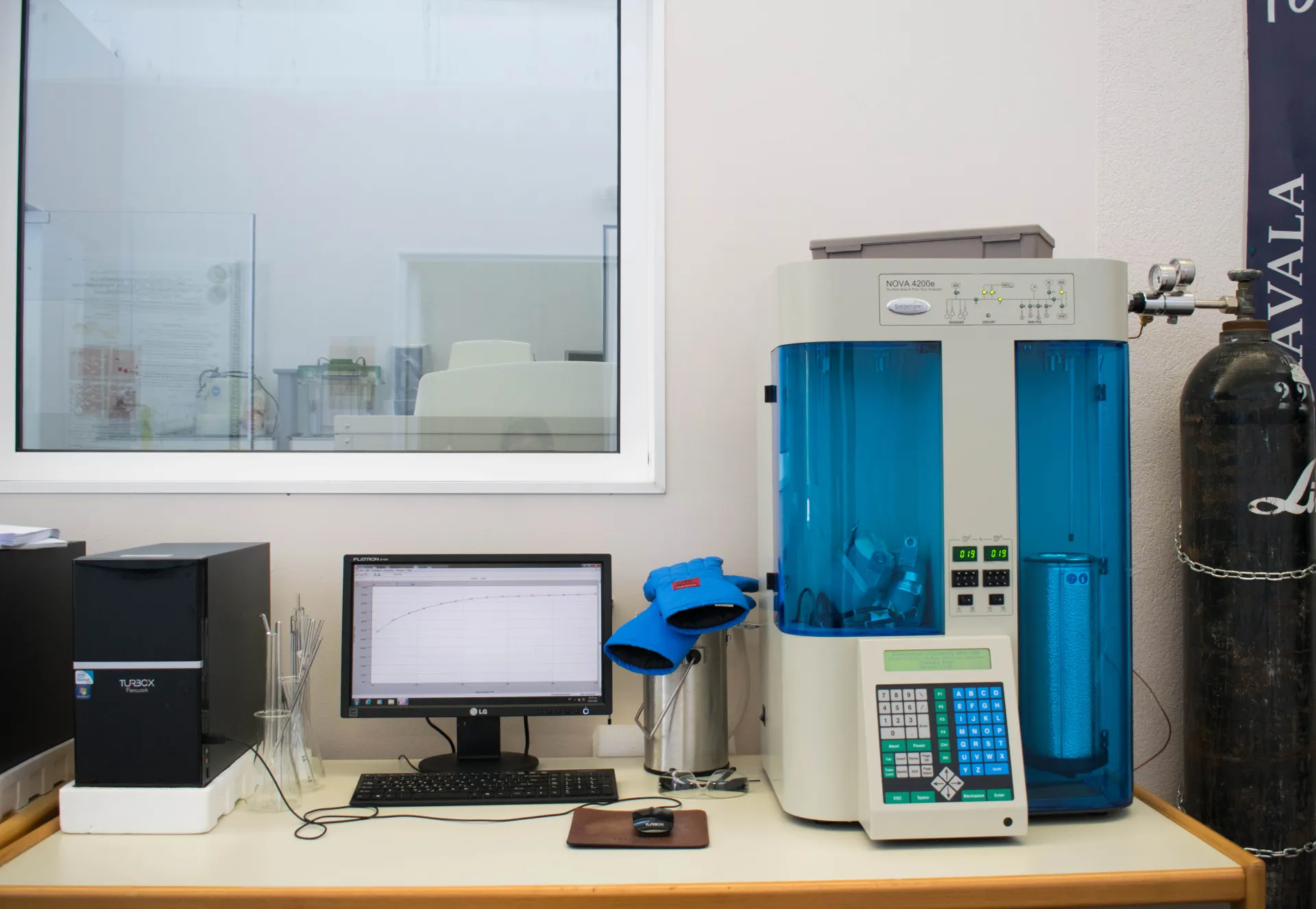
Nitrogen adsorption porosimeter — NOVA 4200e, Quantachrome
Gas adsorption analyzer used to determine specific surface area, pore volume, and pore size distribution of porous materials. Samples are degassed and then exposed to nitrogen at liquid nitrogen temperature to generate adsorption and desorption isotherms. The resulting data can be analyzed using BET theory for surface area and BJH or DFT methods for pore size distribution. The system is suitable for catalysts, activated carbons, ceramics, rocks, cements, pharmaceuticals, battery materials, and other porous solids. Its multi-station configuration allows several samples to be analyzed efficiently.
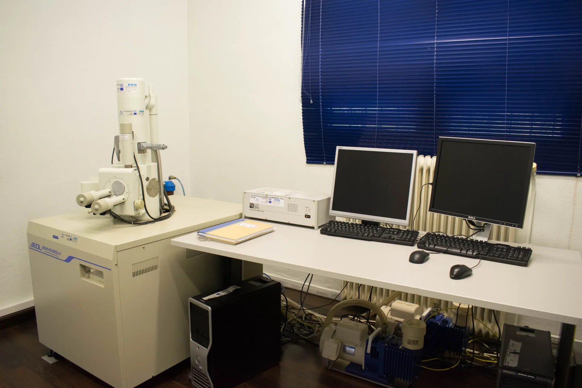
Scanning Electron Microscope — JEOL 6300LV, JEOL
Low-vacuum scanning electron microscope used for high-resolution imaging of sample surfaces and microstructures. A focused electron beam scans the sample, producing signals that reveal surface morphology, texture, particle size, shape, and compositional contrast. The low-vacuum mode allows analysis of non-conductive or partially hydrated samples with reduced charging, while high-vacuum mode supports high-resolution imaging. The SEM is widely used in materials science, geology, biology, electronics, failure analysis, and industrial quality control. It can also support elemental analysis when coupled with EDX.
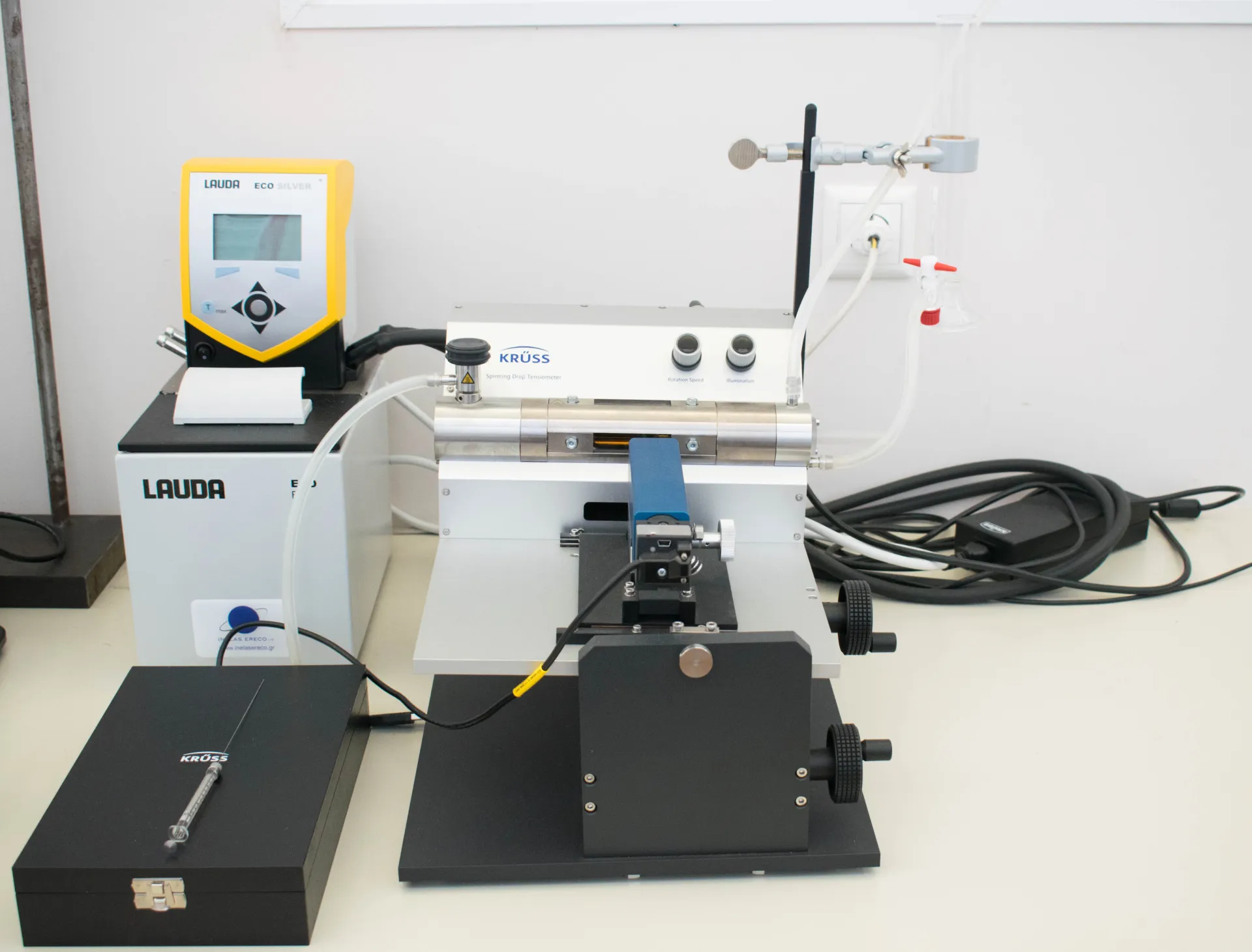
Spinning drop interfacial tension meter — SITE100, Krüss
Instrument for measuring very low interfacial tension between two immiscible liquids using the spinning drop method. A droplet of one liquid is introduced into a capillary filled with another liquid and rotated at high speed. Centrifugal forces elongate the droplet, and imaging software analyzes its shape to calculate interfacial tension. The system is particularly useful for surfactant systems, emulsions, microemulsions, enhanced oil recovery studies, pharmaceutical formulations, cosmetics, and food emulsions where ultra-low interfacial tension values are important.
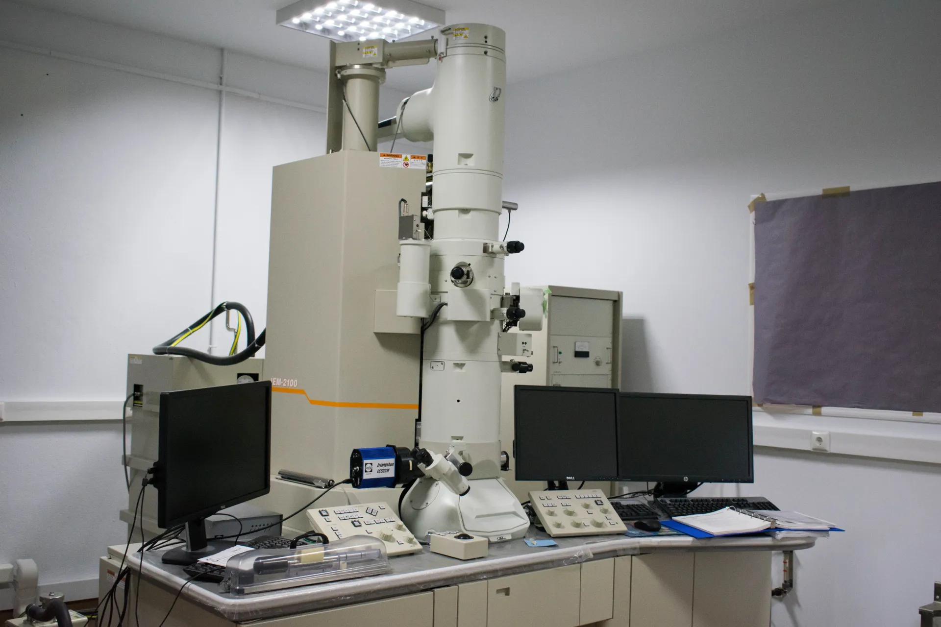
Transmission Electron Microscope — JEOL TEM system, JEOL
High-resolution electron microscope used to examine thin samples at microstructural, nanostructural, and atomic scales. A high-energy electron beam is transmitted through an ultrathin specimen, producing images, diffraction patterns, and analytical signals. TEM enables observation of crystal lattices, nanoparticles, defects, interfaces, phases, and biological ultrastructure. It can be combined with techniques such as electron diffraction, EDX, and EELS for structural and compositional analysis. It is essential for advanced materials science, nanotechnology, chemistry, biology, and semiconductor research.
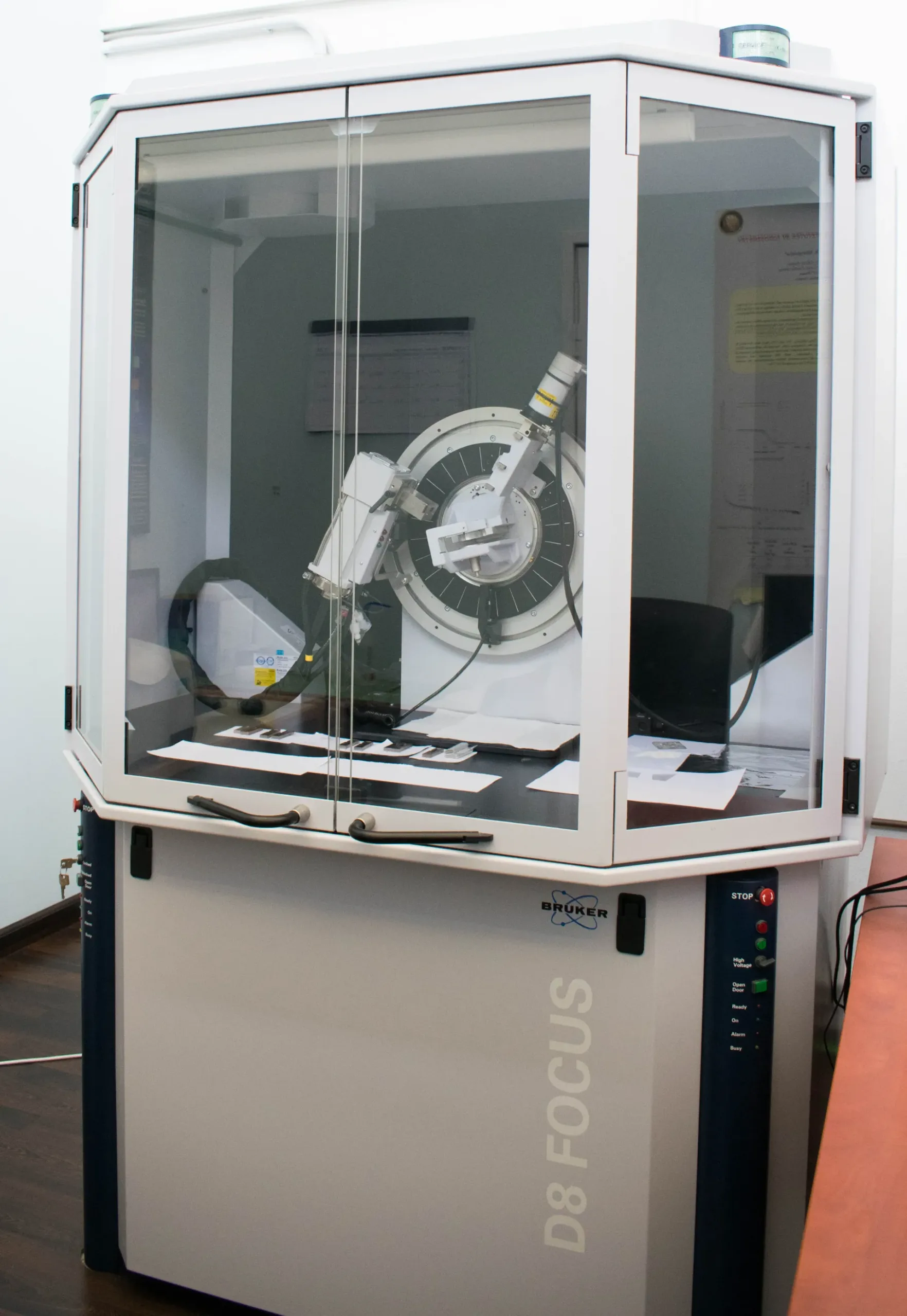
X-ray diffractometer — D8 FOCUS, Bruker
X-ray diffraction system used to determine crystallographic structure, phase composition, lattice parameters, and crystallite size of materials. Powdered samples are exposed to X-rays, and the diffracted intensity is recorded as a function of angle to generate a diffractogram. Peaks in the diffraction pattern correspond to crystal planes and are used for phase identification and structural analysis. The Bruker D8 FOCUS includes a vertical θ/θ goniometer and LynxEye detector for sensitive and rapid data acquisition. It is widely used in materials science, chemistry, mineralogy, pharmaceuticals, and quality control.
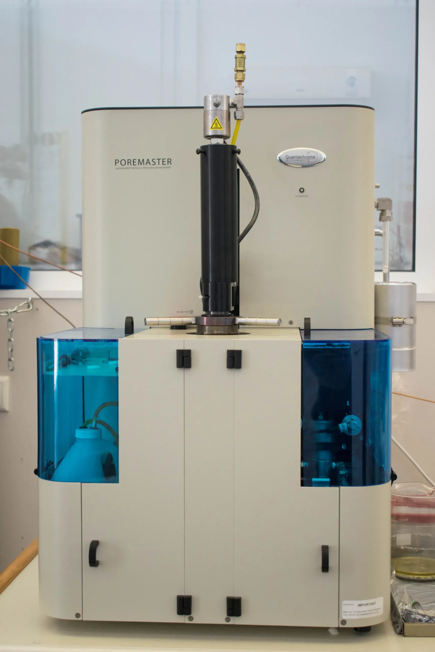
Poremaster Automated Mercury Porosimeter
This advanced mercury intrusion system provides high-resolution analysis of pore size distribution, total pore volume, and macroscopic porosity in solid and powder samples. By applying highly controlled pressure, it enables precise structural characterization, which is essential for advanced research in materials science, nanotechnology, and environmental engineering.